Centre for Microscopy Applied to Biology (CMAbio3)
Contacts
The team
- Head of department: Prof. Jean-Christiophe Avice
- Administrative management
- Guénaelle Levallet (histology platform)
- Benoît Plancoulaine (quantitative histoimaging platform)
- Jean-Christophe Avice (microscopy)
- Technical management:
- Maëlle Guyot (histology platform)
- Nicolas Elie (quantitative histo-imaging platform)
- Didier GOUX (Microscopie)
Activities
The Centre for Microscopy Applied to Biology (CMAbio3) is comprised of three technical platforms:
- A histology platform specialised in the preparation, staining and immunomarking of biological samples. This platform is located within the histology laboratory at the CHU university hospital in Caen.
- A microscopy platform dedicated to the preparation of biological samples for electron microscopy. Observations can be at tissue, cell or subcell level via photonic and electron microscopy. Created in 1968, the platform is located on the Caen University Campus 1.
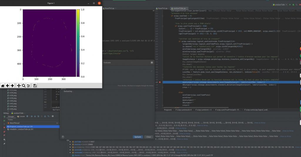
- A quantitative histoimaging platform which offers the CMAbio3 all its originality, for it is specialised in image analysis and processing. This platform is located within the Centre François Baclesse (research building) and at the Caen University Campus 1.
Thanks to the combination of skills and know-how from three technical platforms, the CMAbio3 can now propose a range of complementary services, from technical support to sample handling (cell/tissue), preparation/packaging, staining, morphological or immunochemical analysis, observation (at different resolutions) and computerised image analysis.
The staff of 6 (3 permanent) at the CMAbio3 work together as a team to accomplish their mission to provide services, advice and training for students, technicians, university researchers or external users in all activities: preparation techniques, sample observation and processing/analysis of obtained images. This team work enables the CMAbio3 to offer high added value to research projects involving biological microscopy.
Technical resources
The CMAbio3 is equipped appropriately to allow the preparation, observation and analysis of the biological samples it is entrusted with. CMAbio3 equipment includes:
- Axozoom
- Cryo-ultramicrotome
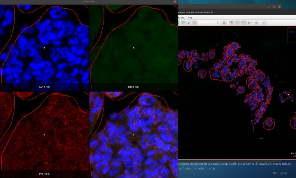
- Confocal laser scanning microscope
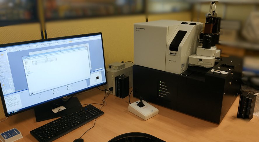
- Scanning Electron Microscope
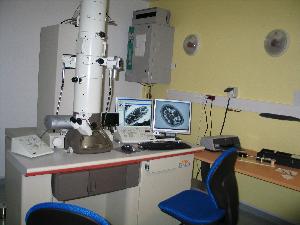
- Transmission Electron Microscope
- Ultramicrotome
- Vibratome
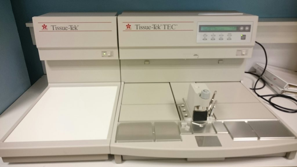
- Paraffin embedding station
- Dako Cryostat
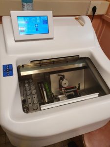
- Routine or special staining automaton
- Immunohistochemistry (IHC)
- Fluorescent hybridation in situ (FISH)
- LASER micro-dissector
- Olympus VS120 slide scanner for brightfield, polarised light and fluorescence digitisation
- Aperio CS slide scanners, computer stations
Partners
At regional level:
- Regional projects with research units from Rouen and Caen
At international level:
- Inter-regional projects and collaborations with European research units