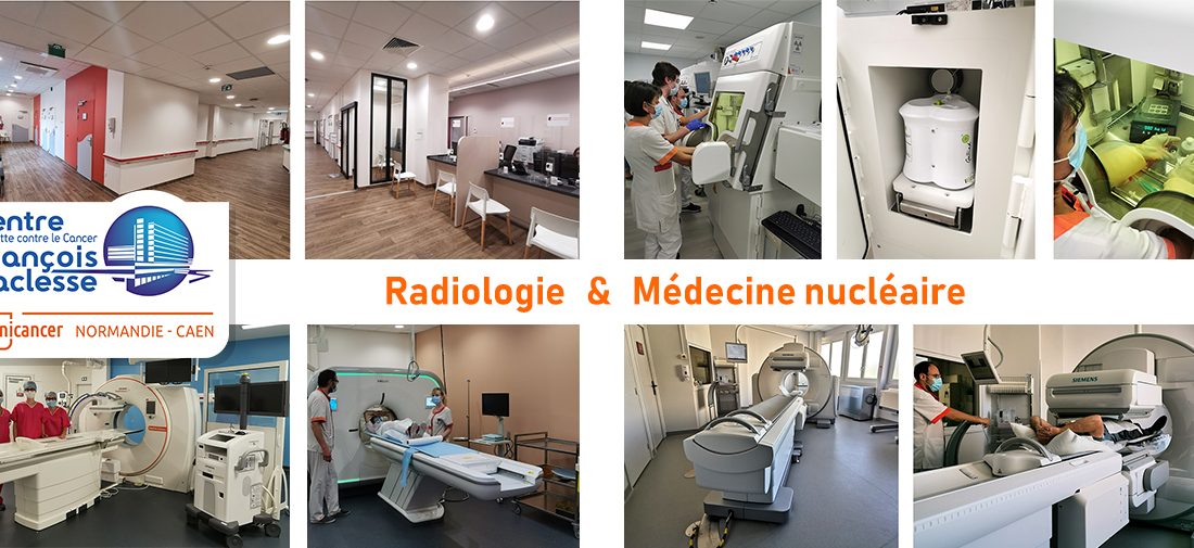
A completely renovated Radiology Department, new state-of-the-art equipment and advanced theranostics
Medical imaging at the Centre François Baclesse has undergone a complete overhaul in recent months. The Radiology department has been completely refurbished, and the Nuclear Medicine department has updated the equipment in its radiopharmaceutical preparation laboratory. In addition, three new state-of-the-art pieces of equipment have been installed (two scanners and a hybrid gamma camera).
What's new in radiology
A new Radiology department to open on June 8, 2020
The Radiology Department is involved at various stages in the care of cancer patients, with diagnostic radiology (to establish a diagnosis, by detecting and characterizing the tumor, its extension and assessing the response to treatment) and interventional radiology (diagnosis and treatment of tumors under radiological guidance, with innovative minimally invasive techniques, without recourse to surgery).
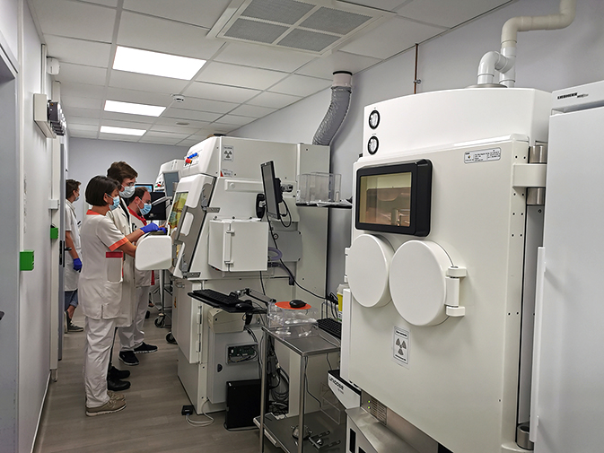
Located on the first floor of the Centre, this department has been completely renovated. It opened after a year and a half's work. During this time, all radiology activities (except MRI) were relocated to other departments at the Center, to ensure continuity of care.
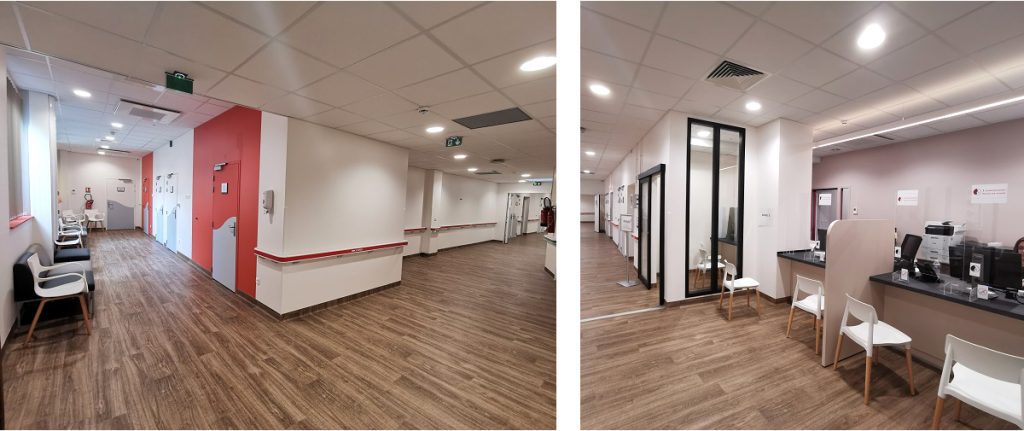
The department has been restructured, becoming more functional, with :
- Improving patient reception,
- Optimizing patient and professional traffic flows,
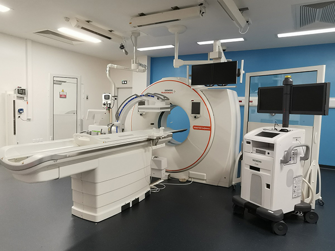
- The development of interventional radiology activities, thanks to the installation of a second scanner, in a controlled environment and under conditions identical to those in an operating theatre.
The new interventional radiology techniques used in this department are :
- Cold treatment of tumors (destruction by cryoablation),
- Hot treatment of tumors (destruction by radiofrequency),
- Bone consolidation by cementoplasty or spondyloplasty.
The radiology department now has a technical platform comprising :
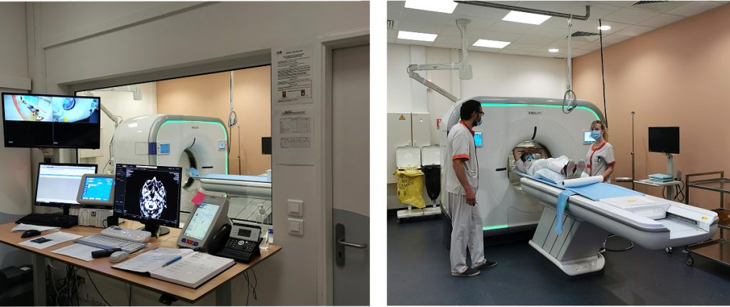
- 1 Philips Incisive CT diagnostic scanner (2020)
- 1 Siemens Confidence RT Pro shared scanner (2020)
- 1 Aéra MRI - Siemens (2017)
- 1 Stephanix Evidence digital X-ray table (2011)
A second scanner, scheduled to open in September 2020
This second scanner, located in the Radiology department, is a Siemens Confidence RT Pro. It will be shared between the Radiology and Radiotherapy departments, with a dedicated activity:
- 30% to interventional radiology,
- 40% to diagnostic radiology: follow-up and management of new patients,
- 30% to radiotherapy, for scans to initiate proton therapy treatment.
(opening in September 2020 under operating theatre conditions)
The opening of this second scanner will enable us to reduce waiting times for scanner appointments, develop our interventional radiology activity, which requires a state-of-the-art scanner, and introduce proton therapy radiotherapy.
And soon, the opening of a new Senology Department in October 2020.
The Senology Unit, which is part of the Radiology Department, is currently being renovated and extended. It will open in October 2020, with new equipment and a new organization.
What's new in Nuclear Medicine
The Nuclear Medicine department carries out diagnostic examinations (scintigraphies and PET scans) and treatments (internal vectorized radiotherapy) using various radioactive products administered to patients. Molecular imaging can be used to explore specific organs, tumors or metabolic pathways.
The Nuclear Medicine department was completely refurbished at the end of 2017, and now boasts a technical platform comprising :
- 1 Vereos Philips digital PET scanner (2017)
- 3 gamma cameras, which detect the gamma rays emitted by radioactive elements and form a scintigraphy (image of their distribution in the body):
- 1 BRIVO GE camera (2016),
- 1 Siemens Symbia T2 camera (2010) coupled with an X-ray scanner,
- 1 new Siemens INTEVO Bold camera coupled with an X-ray tracking scanner (to be installed in June 2020).
- A refurbished radiopharmaceutical preparation laboratory (April 2020)
A new hybrid gamma camera: Symbia INTEVO
This latest-generation gamma camera replaces equipment commissioned in 2006.
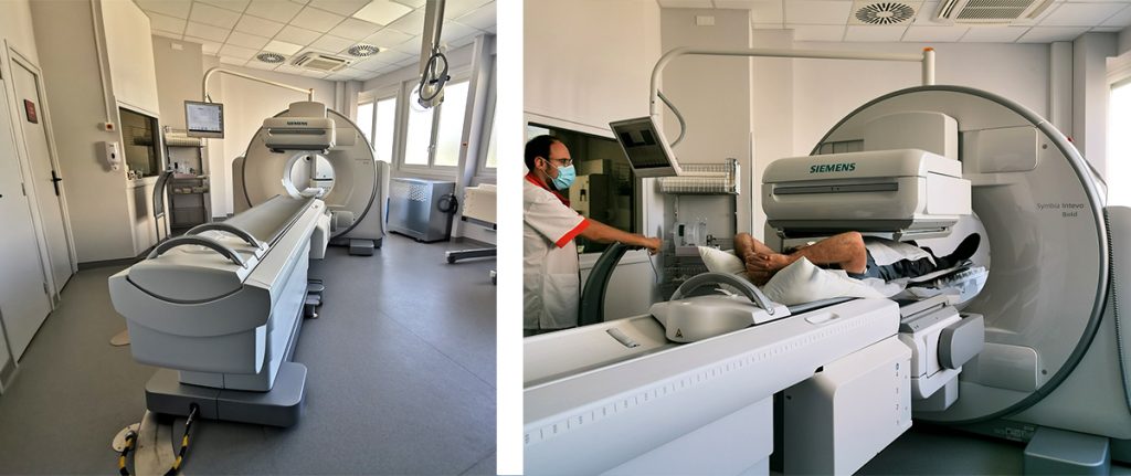
Its main features are :
- Coupling with a high-resolution register scanner (better quality),
- Direct production of very high-definition gamma images, enabling finer interpretations and better visualization of lesions.
- Delivering the lowest possible doses to patients, with faster scans.
A new "hot" radiopharmacy laboratory
In mid-April 2020, a new hot laboratory went into service. Dedicated to the storage, production and control of radiopharmaceutical drugs, it is equipped with :
- two new class A shielded enclosures (also known as glove boxes), enabling radiopharmacists to handle radioelements while protected from radiation. These thicker enclosures enable radiopharmacists to work under excellent radiation protection conditions, while ensuring optimum compliance with hygiene standards when preparing patient doses.
- a Gallium-68 generator for the preparation of new-generation PET-Scan radiotracers, such as DOTATOC (SOMAKIT-TOC ®). This generator has a lifetime of 1 year (March 2020).
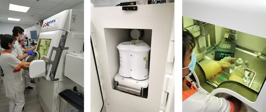
Shielded enclosure / Gallium-68 generator / DOTATOC preparation
A breakthrough in the field of endocrine tumors with the DOTATOC Gallium-68 PET scan and treatment with LUTATHERA
Generally speaking, malignant tumors consume excess glucose, and the tracer usually used for PET scans is FDG, a glucose analogue labelled with a beta + emitter, Fluor 18. This tracer is produced by industrial companies at various cyclotron-equipped sites in France, and delivered to nuclear medicine departments to order.
Some tumors, known as endocrine tumors, do not bind FDG well, but express specific receptors (somatostatin receptors) on their surface.
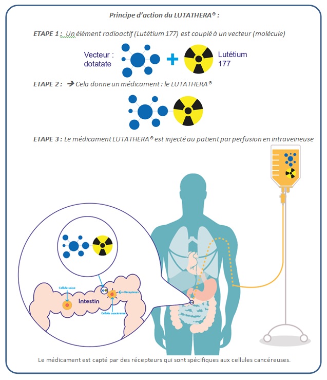
The use of a somatostatin analogue (DOTATOC) labelled with a beta+ emitter, Gallium 68, enables PET images of these lesions, and labelling with a beta-emitter, Lutetium 177, enables treatment (vectorized internal radiotherapy) of these lesions when they are metastatic.
This theranostic pairing ("thera" for therapeutic and "nostic" for diagnostic) represents a major advance in the management of these tumors.
Gallium-68 DOTATOC PET scan
Gallium68 labeling on the DOTATOC vector is carried out in the Nuclear Medicine department, in the "hot" laboratory, inside the shielded enclosures, under the responsibility of a radiopharmacist.
- DOTATOC has a high affinity for somatostatin receptors and binds specifically to endocrine tumors.
- Gallium-68 labelling enables images to be taken on PET scans, with a clear improvement over the Octreoscan scintigraphy used to date.
- The spatial resolution of the PET-Scanner image is much better than that of a gamma camera, with greater detection sensitivity.
- Patient comfort has also been improved, with a DOTATOC PET-Gallium-68 scan lasting between 1h30 and 2h instead of the previous 48h.
Gallium-68 DOTATOC diagnostic tests are used for extension and recurrence assessments. The first patients were treated in April 2020.
Treatment with internal vectorized radiotherapy (LUTATHERA)
This internal vectorized radiotherapy treatment was implemented at the end of 2016 in the Center's 4 dedicated radioprotected rooms on the 7th floor, in patients with metastatic digestive endocrine tumors of the intestine. This treatment has demonstrated its effectiveness and is generally very well tolerated by patients.