Computer-aided 3D (three-dimensional) reconstruction of the mandible (lower jaw) has been available for several years. The complexity of the operation carried out at the Centre François Baclesse was to reconstruct the mandible by combining a titanium prosthesis with a segment of bone taken from another part of the body. The operation, which required the presence of 10 healthcare professionals and lasted over 10 hours, took place on March 30, 2021. Explanations.
Under what circumstances is reconstructive jaw surgery required?
Maxillofacial surgery, one of the components of which is facial reconstruction, can be performed following trauma (gunshot wounds, road accidents, etc.) or, in the majority of cases, following the onset of cancer.At the Centre François Baclesse, patients who benefit from this procedure present :
- or mandibular cancer, requiring surgical removal of the tumor and the jawbone in contact with it,
- or jaw sequelae following cancer treatment. Depending on how advanced the sequelae are, surgery to remove the diseased portion of the mandible may be required.
In these situations, the aim of mandibular reconstruction is to restore bone architecture, so that all jaw-related functions can be maintained: eating, breathing, speaking... To date, the main reconstruction technique involves bone transfer from another part of the patient's body, the "donor".
How can the mandible be reconstructed after cancer?
Reconstructive techniques in maxillofacial surgery have made particular progress since the 1914-1918 war with its famous "broken faces", for whom it was necessary to find a reconstructive solution enabling them to return to a more "normal" life.
For a long time, surgery was very much a craft, with the surgeon's hands and eye being the only elements available to gauge needs. The arrival of new computer-assisted technologies, and in particular the ability to model the patient's jaw in 3D, means that the surgeon can better visualize cutting lines and graft positioning.
To reconstruct the mandible, which defines the lower contour of the face, a segment of bone from the leg or scapula can be harvested (with no significant functional consequences for the harvested limb). The harvested graft is then placed in the area to be reconstructed.
Prior to the operation, the patient undergoes imaging tests that enable 3D modelling of the parts of the body to be operated on.
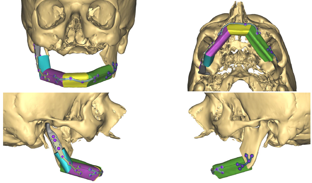
Based on this model, and after defining the area of the jaw to be removed, the surgeon and biomedical engineer simulate the reconstruction of the mandible using the leg bone. 3D-printed cutting guides are designed to ensure precise bone sectioning in both the mandible and fibula. To ensure the strength of the bone reconstruction, 3D-printed titanium plates are fixed and screwed in place.
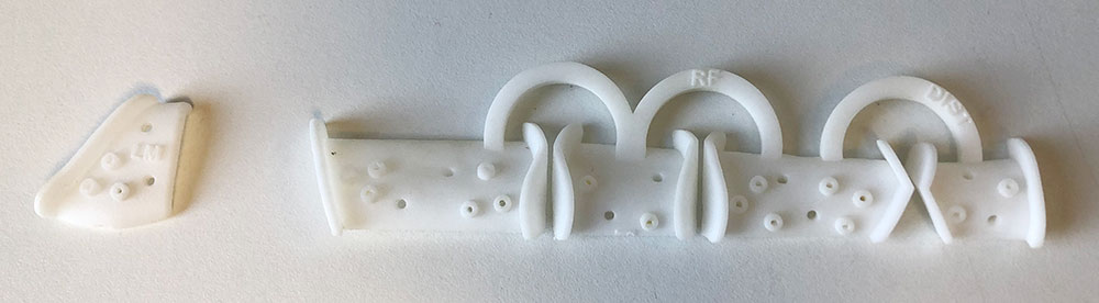
Traditionally, these plates are positioned and modeled manually. In this patient's case, the mandible had to be reconstructed almost entirely, and the fibula harvest was not sufficient to rebuild the jaw. In addition to bone reconstruction, the surgical team had to add a prosthesis. It was this double composition (prosthesis + bone) that added to the difficulty of this first operation at the Centre François Baclesse.
Surgery on the harvested bone is performed at the same time as surgery on the mandible. The surgeons at work perform very delicate and meticulous work to connect the blood vessels. This is microsurgery, with micro-suturing of the vessels. The advantage of autografting is that the graft is not rejected.
Pain management is anticipated. The patient undergoes the procedure under general anesthesia. The anesthetist also performs locoregional anesthesia, in this case on the leg and lower face. Locoregional anesthesia lasts longer than general anesthesia, and reduces post-operative pain.
The benefits of 3D assistance
In addition to the scientific studies already supporting this breakthrough, other studies are seeking to confirm the added value of computer-assisted surgery. Our surgeons can already testify that this technique enables :
- Better functional and aesthetic results,
- Fewer complications, thanks in part to shorter operating times.
The additional cost of this innovative technique, around €2,500 per patient and not reimbursed by health insurance, is fully covered by the Centre François Baclesse, as part of its policy of equal access to excellent care with no extra fees or out-of-pocket expenses for all, without distinction.
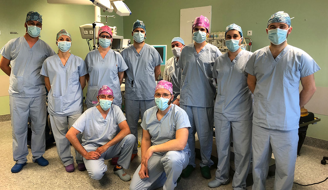
A multidisciplinary team of 10 people at work
The surgeon is often cited as the conductor of this long and delicate operation, but for this first, he worked with a multidisciplinary team of 10 professionals:
- 2 biomedical engineers
- 1 nurse anesthetist
- 1 anaesthetist
- 2 surgeons
- 2 instrumentalists
- 1 surgical intern
- 1 circulating nurse
Dr Julien DROUET, maxillo-facial surgeon at the Centre François Baclesse, trained in Caen and completed his training for 6 months in Switzerland at the Lausanne University Hospital to further his knowledge of computer-assisted surgery. He headed up the operation on March 30.
Dr Audrey LASNE-CARDON, ENT and cervico-facial surgeon, also took part in the operation on March 30.
Improved quality of life after surgery
Between 10 and 15 days after the operation, the patient can resume oral feeding. He will then be fitted with a dental prosthesis, stabilized by implants. This new mandibular architecture, which ensures proper mastication, will enable the patient to regain his or her oral functions, with improved comfort and aesthetics.
The patient will be able to put his or her foot down the day after surgery, and will be able to resume normal activity between 3 and 6 months after the operation.
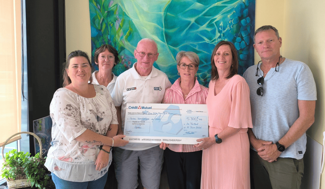
The Centre François Baclesse can treat 4 to 5 patients a year with this technique. It would be possible to offer this treatment to more people, provided that sufficient funds are raised through donations. Donations can be sent to donations.baclesse.fr