The Centre Baclesse and GANIL-LPC teams have developed a scintillation dosimeter to improve the accuracy of proton therapy quality control.
Proton therapy: how does it work?
Proton therapya form of radiotherapy using proton beams, represents a major advance in tumor treatment, particularly for patients requiring optimal protection of healthy tissue. Compared with conventional photon radiotherapy, the use of protons enables a more precise radiation dose to be delivered to the tumor, while minimizing the impact on surrounding tissue.
This enhanced precision is further enhanced by the ability of proton beams to be very thin and to operate in pulses, optimizing dose adaptation to tumor contours . This makes proton therapy an invaluable tool for treating certain complex and delicate tumors, where other forms of radiotherapy could lead to significant side effects.
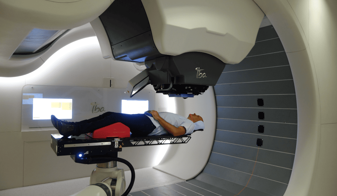
Before treatment, it's important to check that the fine pulsed beams deliver the right dose, at the right position, with the right energy and intensity. This is the job of the medical physicists who check all this for each patient (quality control stage) at Cyclhad, the proton therapy center in Normandy. But this is a time-consuming and difficult task in the clinic, as the equipment used for these measurements (commercial dosimeters for proton therapy), is not always sufficiently accurate (reduced spatial and temporal resolution).
Dans ce contexte, un dosimètre à scintillation (nommé SCICOPRO) permettrait une très bonne résolution spatiale (< 1 mm) et des mesures en 3 dimensions.
Working together
The scintillation dosimeter was developed in collaboration with GANIL, the Caen Corpuscular Physics Laboratory and the Centre Baclesse. This PMRT project (Plateforme de Modélisation en Radiothérapie) was funded by a call for projects from the Normandy Region and the European Union.
The system (Figure 1 A) can emit visible light (scintillation) when irradiated. An ultra-fast CMOS camera records the scintillation produced by each pulse, and all the pulsed beams are used to reconstruct the dose.
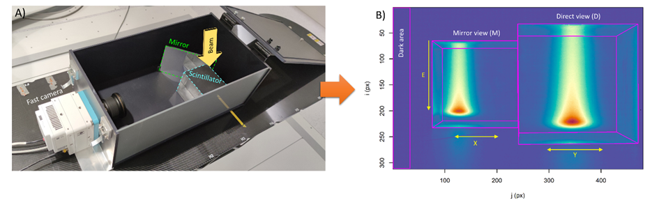
B) Typical image acquired when the cube is irradiated with vertical proton beams. The camera records a direct view of the pulse on the right-hand side of the image and a view reflected by the mirror on the left-hand side of the image.
This prototype dosimeter is capable of measuring all the characteristics of pulsed fine beams in a single acquisition, with an accuracy of 580 µm for position, 180 keV for energy and 3% for intensity.
This performance is very promising and meets the needs of clinical practice. The dosimeter can detect orientation errors as small as 1°. The prototype is currently being improved to calculate dose distribution in 3 dimensions.
- A publication appeared in the reference journal of medical physics: Frelin AM*, Daviau G*, Bui MHH*, Fontbonne C°, Fontbonne JM°, Lebhertz D, Mainguy E*, Moignier C, Thariat J, Vela A. Development of a three-dimensional scintillation detector for pencil beam verification in proton therapy patient-specific quality assurance. Med Phys. 2024 Sep 10. doi: 10.1002/mp.17388.
The aim of the PMRT (Radiotherapy Modeling Platform) is to collect comprehensive data on the treatments applied (external radiotherapy using X-rays, protons and hadrons) and the clinical results of these treatments, in order to develop models for tumor control and, above all, for the induction of side effects.