The APACH 2 study conducted by a team of researchers from the Baclesse Center has just been published on the front page of the prestigious international medical journal JAMA Otolaryngology–Head & Neck Surgery. This study compared two medical imaging techniques in the surgical management of primary hyperparathyroidism.
What is hyperparathyroidism?
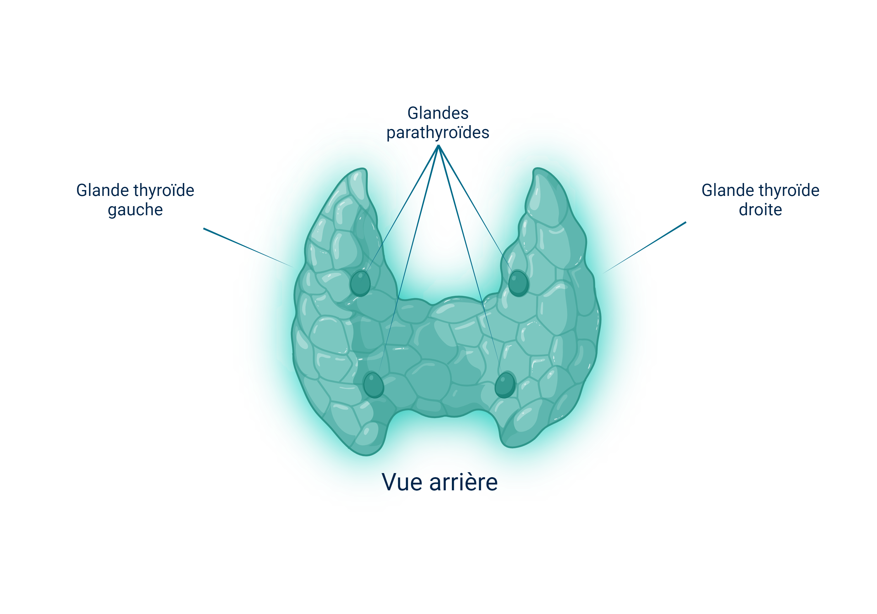
The parathyroids are 4 small glands, about the size of a grain of rice , located in the lower neck to the right and left of the thyroid. They produce parathyroid hormone, which regulates calcium levels in the blood.
Sometimes, a small benign tumor called a parathyroid adenoma develops on one of the parathyroids. The adenoma increases blood calcium levels (hypercalcemia) through excessive production of parathyroid hormone. This condition is known as primary hyperparathyroidism.

Which treatment?
Hypercalcemia is a health risk, which is why surgical removal of the adenoma is indicated.
The advantages of this minimally invasive surgery are :
- Its short life
- Its efficiency
- Lower risk of complications
- Reduced scar size
But how do you know which parathyroid has an adenoma?
Thanks to technological advances in medical imaging, surgical exploration of 4 parathyroid sites has gradually been replaced by so-called "minimally invasive" surgery performed on an outpatient basis. When imaging enables the adenoma to be located, the surgical procedure can be targeted and minimally invasive. On the other hand, when imaging fails to localize the adenoma, more extensive surgery exploring all 4 sites is performed.
Fluorocholine PET scan, a precision technology
The latest technological advance for visualizing parathyroid adenoma in more patients and with greater precision is the fluorocholine PET scan.
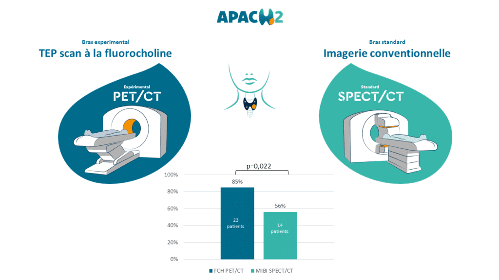
The APACH 2 clinical trial was conducted in collaboration with teams in Brest and Rennes, involving 57 patients with primary hyperparathyroidism, who were randomly assigned to undergo either fluorocholine PET scan or conventional imaging (= Tc99m-sestaMIBI scintigraphy).
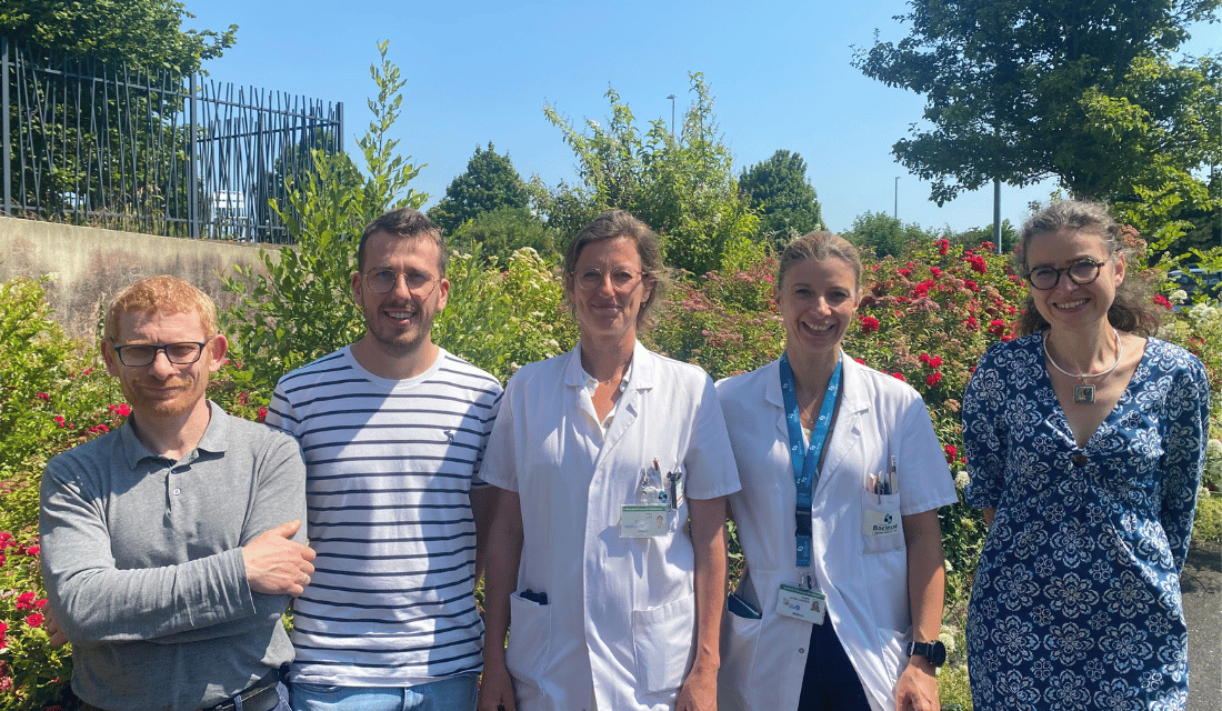
The members of the Centre Baclesse team (see photo at the beginning of the article) who worked on the study are :
- Jean-Michel GRELLARD (Clinical Research Project Manager)
- François CHRISTY (Clinical Research biostatistician)
- Dr Elske QUAK (nuclear physician and principal investigator)
- Dr Audrey LASNE-CARDON (ENT and cervico-facial surgeon)
- Bénédicte CLARISSE (Clinical Research Promotion Pharmacist)
It has been shown that first-line PET scanning enables 85% of patients to benefit from minimally invasive surgery followed by normalization of blood calcium levels, compared with 56% of patients who underwent conventional imaging. The ability of PET scans to accurately locate parathyroid adenomas, i.e. the sensitivity of the examination, was 82% for PET scans versus 63% for conventional imaging. In addition, no adverse events related to the fluorocholine PET scan were reported.
The conclusion of this study is therefore that fluorocholine PET scan can effectively replace conventional imaging to guide the type of surgery for patients with primary hyperparathyroidism. Fluorocholine PET scans could enable more patients to benefit from minimally invasive surgical resection of parathyroid adenoma, resulting in normalization of blood calcium levels.