The Baclesse Center is launching its own artificial intelligence (AI) hub to bring together the various work carried out by its researchers. The goal? To create a structure that unites the Center's various professionals working on AI. The fields of activity are numerous and include medical imaging, radiotherapy, brachytherapy, anatomic pathology, and the patient care pathway. Artificial intelligence is currently being developed in many fields of application, including mathematics, statistics, probability, and computer science. Its goal is to enable machines to imitate human intelligence through the creation and application of algorithms. This approach is comparable to the creation of a gigantic "tree" of instructions, applied to the field of medical research.
A tool at the heart of medical research
In the healthcare field, its potential is of interest to researchers and healthcare professionals alike. Artificial intelligence brings many benefits to patients by improving diagnostic accuracy, personalizing treatments, enhancing care management and enabling more efficient health monitoring. To achieve this, algorithms need access to large and varied data sets to train themselves, improve their accuracy and eliminate bias.
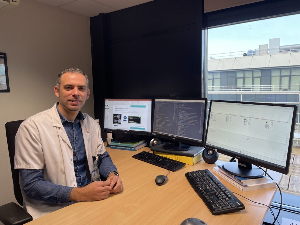
It was this possibility that interested the head of this AI division, Aurélien Corroyer-Dulmont, a researcher and engineer in medical imaging. He is also part of the ISTCT research unit "Imaging and Therapeutic Strategies for Cancer and Brain Tissue" led by Dr. Myriam Bernaudin and hosted on the Cyceron platform. Through various projects carried out at the Center since 2019, he has been able to explore possible applications for AI.
Improved appointment times for PET scans
This AI cluster has two objectives: to evaluate AI solutions for subsequent use, and to develop in-house solutions to meet specific needs. Several tools have already been tested at the Center, notably in medical imaging. Thanks to a research project carried out on a specific type of imaging, PET (Positron Emission Tomography), the acquisition of images has been considerably accelerated. The positive results of this study enabled the system to be used on a routine basis. As a result, the service has gone from 18 to 25 patients a day, which represents a clear improvement in appointment times. Evaluating this type of tool has also enabled us to understand how the algorithms used work, so that we can develop new ones.
A similar approach was taken in the radiology department for MRI imaging. The article validating this approach will be published in 2024 inCancer et Radiothérapie (Lemaire et al., 2024).
PET (Positron Emission Tomography) is an advanced medical imaging technique used to visualize the inside of the human body. It is mainly used to detect abnormalities or diseases in tissues and organs. It provides detailed information on the body's inner workings, helping doctors to plan appropriate treatments. This examination is carried out in the nuclear medicine department of the Centre François Baclesse.
Brain tumors: towards AI prediction of treatment efficacy
A new project starts in November 2023, with the science thesis of Noémie Moreau (supervised by Aurélien Corroyer-Dulmont), a PhD student at the Centre François Baclesse funded 50% by the Normandy region. This project, carried out in collaboration with radiotherapists and radiologists at the Centre François Baclesse, investigates the possibility of AI prediction of treatment efficacy. In this case, the team will be looking at the treatment of brain tumors (glioblastomas, meningiomas and brain metastases) by radiotherapy. The aim is that, thanks to data stored in medical imaging, AI will be able to predict the response to treatment even before it has begun, leading to real personalization of care. In particular, this will make it possible to avoid treatments and thus unnecessary side effects, in cases where it is known that the patient will not respond to them. The Center's teams working on AI-related research projects collaborate closely with GREYC (Groupe de Recherche en Informatique, Image et Instrumentation de Caen). The GREYC laboratory carries out research activities in the field of digital sciences. It has 180 members and is made up of 7 research teams with teacher-researchers fromENSICAEN andUNICAEN, CNRS researchers, PhD students and administrative and technical staff.
France's first federated apprenticeship
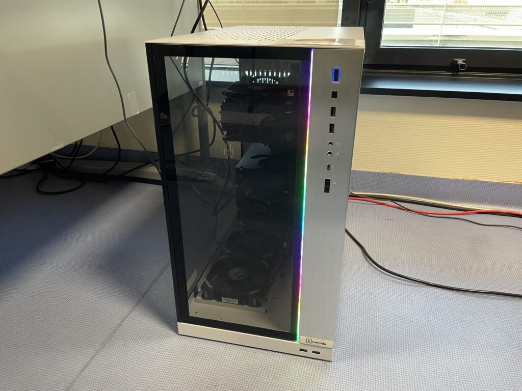
It was in response to the Normandy Region's call for projects,"Booster IA 2022,"that Aurélien Corroyer Dulmont and Romain Modzelewski (Biomedical IT Manager at the Henri-Becquerel Cancer Center in Rouen) were able to develop a joint project around AI and purchase a computing station for the development of artificial intelligence algorithms.
The main limitation of AI algorithms is their use outside the hospital where they were trained. Whatever the problem, an algorithm will depend on the type of data used for training. Thus, using the algorithm in a hospital with different data will render the model much less effective.
"There could be an analogy when, for example, we teach a very young child to recognize apples among a selection of fruits, but with only one variety of apple. If, after this learning process, we present them with an apple of a different variety (different size, shape, or color), they will be confused, if not completely lost. However, it is obviously desirable that a solution developed in one center can be used by others. This is a necessity for it to be useful in practice,"explains Aurélien Corroyer-Dulmont.
That's why Aurélien Corroyer Dulmont and Romain Modzelewski have developed a project for federated learning of artificial intelligence algorithms between hospital sites in Normandy. The aim is to demonstrate that federated learning delivers better results faster, and to acquire the skills and methodology to implement this type of federated project on a larger scale.
Care pathways and quality
AI can also helpimprove the quality of care by bringing out new quality criteria in patient care.
The aim? To use the potential of AI to optimize the patient care pathway by highlighting currently non-visible quality-of-care indices that would enable important variables in patient management to be better taken into account. Analysis of these criteria could have an impact on patients' recurrence-free survival.
Dr Lawrence Nadin, an IMG physician at the Centre Baclesse, began a thesis in September 2023 based on retrospective data on breast cancer patients. The DIM physician is responsible for health data collection and coding quality, and ensures the confidentiality of patient data within the establishment.
In collaboration with the Inserm Anticipe research unit and the MapInMed platform, Dr Nadin is analyzing the relationship between the socio-economic and territorial environment of breast cancer patients, and the quality of their care pathway. To do so, he relies on the identification of lower compliance with IQSS (Indicateurs de la Qualité et de la Sécurité des Soins) as defined byINCa (Institut National du Cancer), longer treatment delays, and a higher propensity to present atypical care pathways. In particular, they concern the interval between the warning mammogram and the first treatment, the use of radiotherapy after conservative surgery, and the time between surgery and the first complementary treatment.
The question is simple: are there identifiable criteria at the start of the care pathway that would enable cancer care professionals to be more attentive to certain patient profiles at risk of deviating from the standard care pathway?
This project has multiple benefits. For patients, it is possible to mitigate unfavorable socioeconomic and regional circumstances with "facilitators" such as therapeutic education, care nurses, or social workers. Adherence to and access to care will thus be improved in unfavorable circumstances. For healthcare institutions, having these predictive criteria will facilitate patient care and ensure equal access to treatment.
AI and anatomo-pathology: a finer work than the doctor's eye?
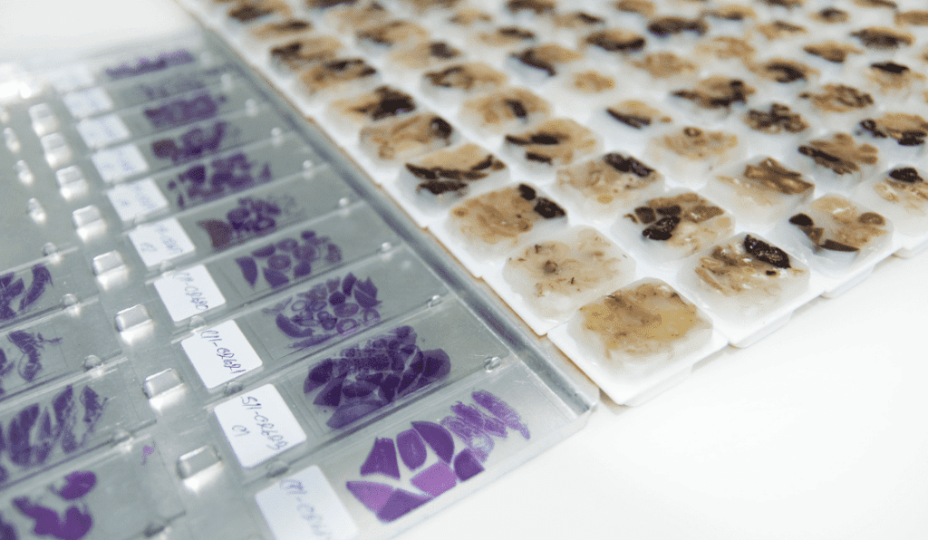
As part of the C³ cooperative project "IAIN: Artificial Intelligence, Digital Imaging," the François Baclesse (Caen), Oscar Lambret (Lille), and Henri Becquerel (Rouen) Centers have acquired slide scanners and opted for a single, shared image management solution.
All pathology samples are preserved in small kerosene blocks. A thin slice a few microns thick (ribbons) is made from each block, placed on a glass slide and stained for microscopic examination. The standard pathology stain is HES (Hemalun-Eosin-Safran). This stain combines hemalun, which stains nuclei purple, eosin, which stains cytoplasm pink, and saffron, which stains collagen fibers yellow. Complementary stains may also be used for certain types of tumour.
This project will enable anatomopathologists to share slides instantly with colleagues from other hospitals to discuss difficult cases, develop image analysis tools to improve diagnosis and research projects to decipher tumour lesions. This cooperation is also a way of tackling the problem of unfavorable medical demographics, by pooling technical resources to improve data exchange. Artificial intelligence will then be able to support the pathologist's work with a tool capable of providing a finer quantitative analysis of complementary stains than the human eye.
Installation of these new blade scanners is scheduled for 2024.
New research on the horizon
The creation of this AI cluster will facilitate the development of AI-related projects in the fields in which we work. It opens up new perspectives in the field of medical research and places the Centre François Baclesse at the heart of scientific advances.
AI and 3D printing
The H3DMED research project, led by the François Baclesse Center, received €246,000 in ERDF funding as part of the Normandy Region's call for projects on "Data and related technologies applied to health." Launched in June 2021 and carried out in partnership with Evanov SAS (Caen), this project has demonstrated that personalized brachytherapy treatments for skin and gynecological cancers have the potential to be optimized and used in healthcare facilities through the combined use of artificial intelligence and 3D printing.